| Cleared and Stained Cichlids |
| Cleared and Stained Cichlids |
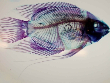
Clearing and Staining allows scientists to view the internal parts of a cichlid intact. Bones stain red while cartilage stains blue. Unlike an X-ray, which is an image of the fish, this technique actually digests away the soft tissues to leave the hard tissues in place for permanent study. Scientists can then study how the various bones and cartilages connect.
I am indebted to Tom Waltzek (Section of Evolution and Ecology, University of California, Davis) for the images on this page. Tom is doing research on the feeding morphology of various cichlids in the lab of Dr. Peter Wainwright.
|
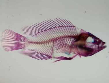
Caquetaia spectabilis. This specimen was cleared and stained by Tom Waltzek of the University of California, Davis. |
|
|
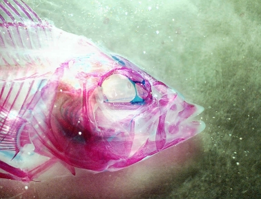
Closeup of the head of Caquetaia spectabilis. Notice the complex bone structure in the jaw area. Like several other cichlids, this species is able to rapidly protrude its jaw to capture prey. The large spaces in the head region hint at this ability. This specimen was cleared and stained by Tom Waltzek of the University of California, Davis. |
|
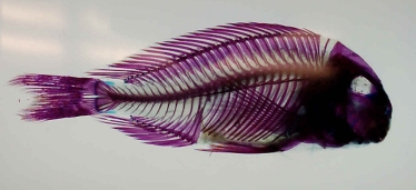
Tropheus polli. Compare this specimen with the one above. Notice the density of the bones in the head region. This species relies on strength in the mouth region to crush and grind its food. This specimen was cleared and stained by Tom Waltzek of the University of California, Davis. |
|
|
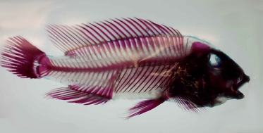
Neolamprologus tretocephalus. This specimen was cleared and stained by Tom Waltzek of the University of California, Davis. |
|
Mesonauta festivum. Commonly called the festivum, in life this fish looks quite different from other cichlids because of its color pattern, a slanted dark stripe. Cleared and stained we see that it is in fact much like other South American cichlids. This specimen was cleared and stained by Tom Waltzek of the University of California, Davis. |
|
![[Go Back to Contents]](gifs/back.gif) |
![[Send email to Ron Coleman]](gifs/address.gif) |